

Plate I. Right hand of the anatomist, dorsal view, holding nothing. Note the callus on the middle finger, where the scalpel rested for thirty-eight years. Note the tremor, which a plate cannot show. Imagine it.
Plate II. Everything has a name. Hunter’s canal. Wharton’s duct. The circle of Willis, which Willis did not discover and wore anyway, like a borrowed coat. The anatomist has checked. Nothing in the body is still open. The last unclaimed structure was taken before he was born.
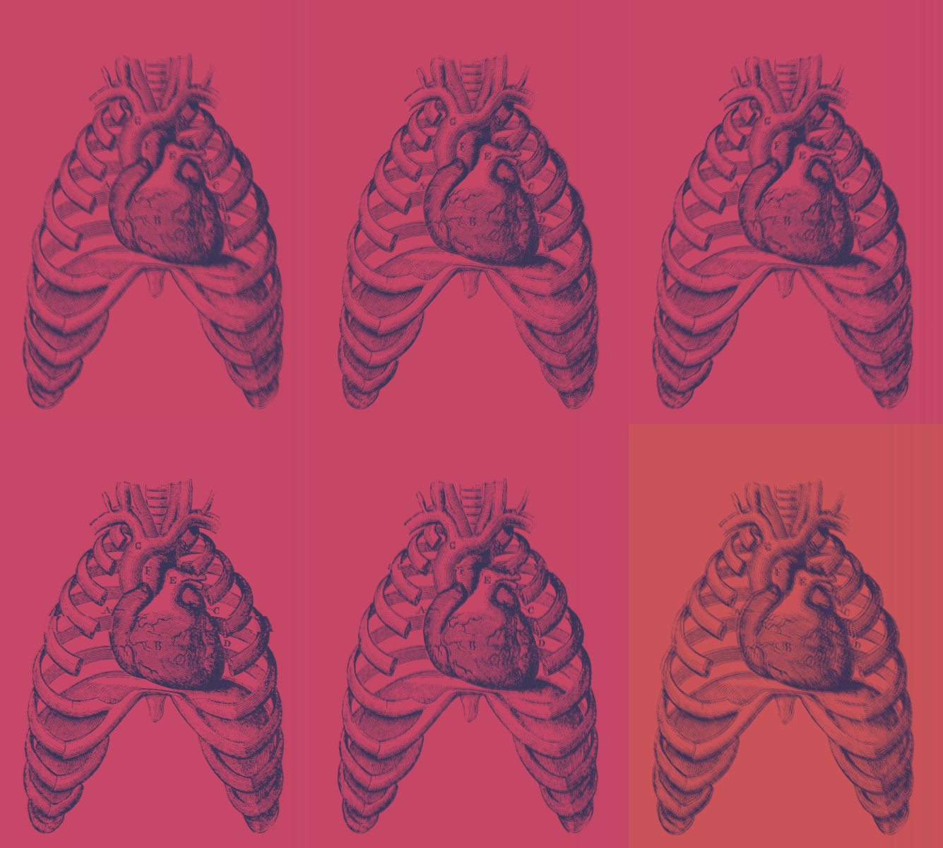
Plate III. Anterior mediastinum, donor 114, female, 81. Between the posterior sternum and the pericardium, a space the size of a thumbprint. Not the thymic remnant. The thymic remnant is labeled (a). This is (b). It has no fat, no vessels, no innervation. It has edges. The anatomist presses a probe into it, and the probe goes in farther than the chest is deep.
Plate IV. Blank. The engraver declined.
Plate V. Donors 115 through 131. (b) present in all. In donor 122, a retired letter carrier, the space contains a single folded page. The anatomist does not unfold it. Protocol requires all tissue be returned to the family for cremation. He does not know whether a page is tissue. He decides it is.
Plate VI. The anatomist’s wife at the kitchen table, lateral view. She is saying something. Note the orbicularis oris, which is doing the work. Note the anatomist’s hand reaching for the salt, which is the only answer he gives.
Plate VII. Proposed nomenclature: fossa retrosternalis. Fossa of Drury. The committee returns the letter. Enclosed: a reminder that one may not name what one has not published, and one may not publish what cannot be reproduced, and when the committee opened its own donors it found (a), and it found the pericardium, and it found nothing between.
Plate VIII. Donor 132. The anatomist’s hands, which have never trembled in the lab, tremble. He has read the intake card. He did not know she had signed the form. She signed it in 2009, the year of the salt.
Plate IX. Omitted at the request of the family, which is the anatomist.
Plate X. Anterior mediastinum, donor 132. (b) is present, larger than in any prior specimen. The anatomist inserts the probe and does not find the bottom. He inserts his finger and does not find the bottom. The students, who have been told to observe, observe.
Plate XI. The students’ notes, compiled. Most write (b). One draws the anatomist’s arm entering the chest to the elbow and labels it correctly: brachioradialis, flexor carpi ulnaris, and so forth. It is the best drawing in the class. He gives it full marks and never learns her name.
Plate XII. Right hand of the anatomist, dorsal view, closed around a folded page. Compare Plate I. Compare Plate V. Do not unfold it.